Catalogue

Recombinant mouse anti vimentin
Catalog number: MUB1903P| Clone | RV204 |
| Isotype | IgG1 |
| Product Type |
Monoclonal Antibody |
| Units | 0.1 mg |
| Host | Mouse |
| Species Reactivity |
Human Mouse |
| Application |
Immunoblotting Immunocytochemistry Immunohistochemistry (frozen & paraffin) |
Background
Vimentin (57 kDa) is the intermediate filament protein (IFP) of mesenchymal cells. This IFP however often deviates from the tissue-specific and developmentally regulated pattern of expression. Besides its typical expression in most cultured cells, vimentin is also expressed together with several other IFPs during early stages of development. As differentiation proceeds, vimentin is exchanged for the tissue-specific intermediate filament type. Also in cancers, vimentin is often expressed in addition to the tissue-specific IFP.
Source
MUB1903P (clone RV204) is a recombinant mouse anti vimentin monoclonal antibody produced in mammalian HEK293 cells.
Clone RV204 is produced by cloning the antibody sequence of the mouse anti vimentin hybridoma RV202, which was derived by fusion of SP2/0-Ag14 mouse myeloma cells with spleen cells from a BALB/c mouse immunized with a cytoskeletal vimentin extract of calf lens.
Product
Each vial contains 100 ul 1 mg/ml ProtA purified recombinant monoclonal antibody in PBS containing 0.09% sodium azide.
The purity of the antibody is >98% as determined by SDS-Polyacrylamide gel electrophoresis.
The endotoxin level is < 1EU/mg as determined by the LAL chromogenic endotoxin assay.
Concentration: 1 mg/mL
Specificity
Human and mouse vimentin.
Pieper et al (European Journal of Biochemistry 1992; included) show in figure 5 and 6 that replacement of exons 7-9 in the vimentin gene (construct Vvim2) results in a vimentin protein that is no longer detected by RV202, indicating that the epitope for RV202 is located in the C-terminal region of vimentin, comprising amino acid range 409-466 .
Species Reactivity: MUB1903P (clone RV204) reacts exclusively with vimentin, which is expressed in mesenchymal cells and mesenchymal derived tumors e.g. lymphoma, sarcoma and melanoma. When applied to either frozen or paraffin sections of human tissues the antibody reacted with the appropriate tissues and cells, such as endothelia, vessels, stellate cells, glomerular podocytes, nerves, vessels, myoepithelial cells, adipocytes, endometrial glands, and Sertoli cells, with no staining in epithelial cells.
Applications
MUB1903P (clone RV204) is suitable for immunoblotting, immunocytochemistry, and immunohistochemistry on frozen and paraffin-embedd tissue sections. Optimal antibody dilution should be determined by titration; recommended range is 1:100 – 1:200 for immunohistochemistry with fluorochrome conjugated secondary antibodies or with avidin-biotinylated horseradish peroxidase complex (ABC) as detection reagent, and 1:100 – 1:1000 for immunoblotting applications.
Storage
The antibody is shipped at ambient temperature and may be stored at +4°C. For prolonged storage prepare appropriate aliquots and store at or below -20°C. Prior to use, an aliquot is thawed slowly in the dark at ambient temperature, spun down again and used to prepare working dilutions by adding sterile phosphate buffered saline (PBS, pH 7.2). Repeated thawing and freezing should be avoided. Working dilutions should be stored at +4°C, not refrozen, and preferably used the same day. If a slight precipitation occurs upon storage, this should be removed by centrifugation. It will not affect the performance or the concentration of the product.
Shipping Conditions: At ambient temperature
Caution
This product is intended FOR RESEARCH USE ONLY, and FOR TESTS IN VITRO, not for use in diagnostic or therapeutic procedures involving humans or animals. It may contain hazardous ingredients. Please refer to the Safety Data Sheets (SDS) for additional information and proper handling procedures. Dispose product remainders according to local regulations.This datasheet is as accurate as reasonably achievable, but Nordic-MUbio accepts no liability for any inaccuracies or omissions in this information.
Safety Datasheet(s) for this product:
| NM_Sodium Azide |
|
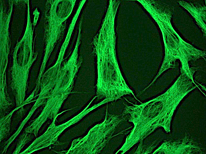
Figure 1. Indirect immunofluorescence staining of normal human dermal fibroblasts in tissue culture with MUB1903P (diluted 1:250), showing the specific cytoskeletal pattern of vimentin intermediate filaments. |
|
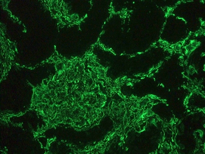
Figure 2. Indirect immunofluorescence staining of human kidney tissue section with MUB1903P (diluted 1:1000), showing the specific pattern of vimentin in the mesenchymal cell types, such as fibroblasts in the connective tissue, podocytes, and endothelial cells in blood vessels. As expected, no reactivity is seen in the epithelial cell compartment |
|
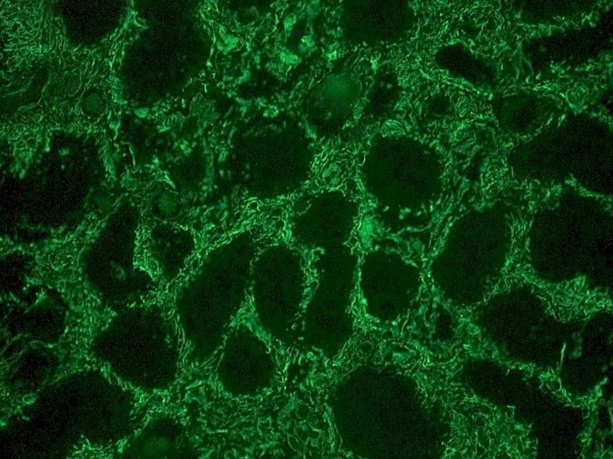
Figure 3. Indirect immunofluorescence staining of human kidney tissue section with MUB1903P (diluted 1:1000), showing the specific pattern of vimentin in the mesenchymal cell types, such as fibroblasts in the connective tissue, podocytes, and endothelial cells in blood vessels. As expected, no reactivity is seen in the epithelial cell compartment |
|
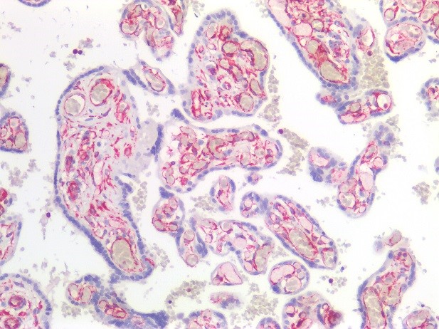
Figure 4. Immunostaining of human paraffin embedded tissue section of placenta with MUB1903P (diluted 1:100), showing the specific pattern of vimentin in the mesenchymal cell types, such as fibroblasts in the connective tissue, and endothelial cells in blood vessels. As expected, no reactivity is seen in the epithelial cell compartment |
|
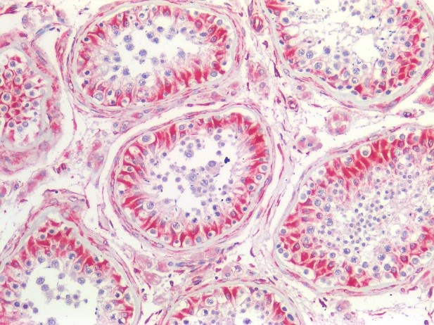
Figure 5. Immunostaining of human paraffin embedded tissue section of testis with MUB1903P (diluted 1:100), showing the specific pattern of vimentin in the mesenchymal cell types, such as fibroblasts in the connective tissue, endothelial cells in blood vessels and Sertoli cells. |
|
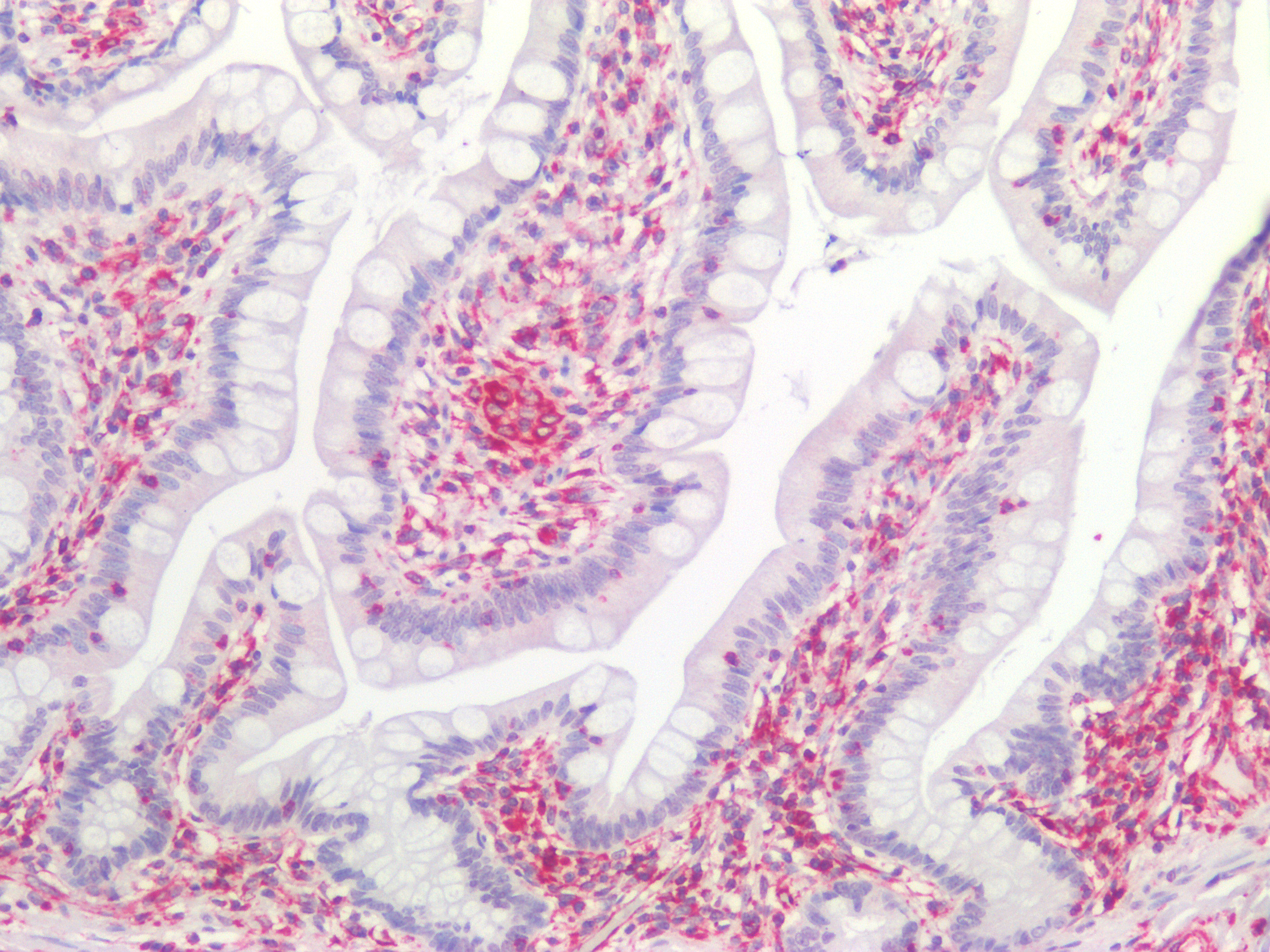
Figure 6. Immunostaining of human paraffin embedded tissue sections of human colon with MUB1903P (diluted 1:100), showing the specific pattern of vimentin in the mesenchymal cell types, such as fibroblasts in the connective tissue, and endothelial cells in blood vessels. As expected, no reactivity is seen in the epithelial cell compartment. |

|
Figure 7. Immunostaining of human paraffin embedded tissue sections of human kidney with MUB1903P (diluted 1:100), showing the specific pattern of vimentin in the mesenchymal cell types, such as fibroblasts in the connective tissue, and podocytes. As expected, no reactivity is seen in the epithelial cell compartment. |
|
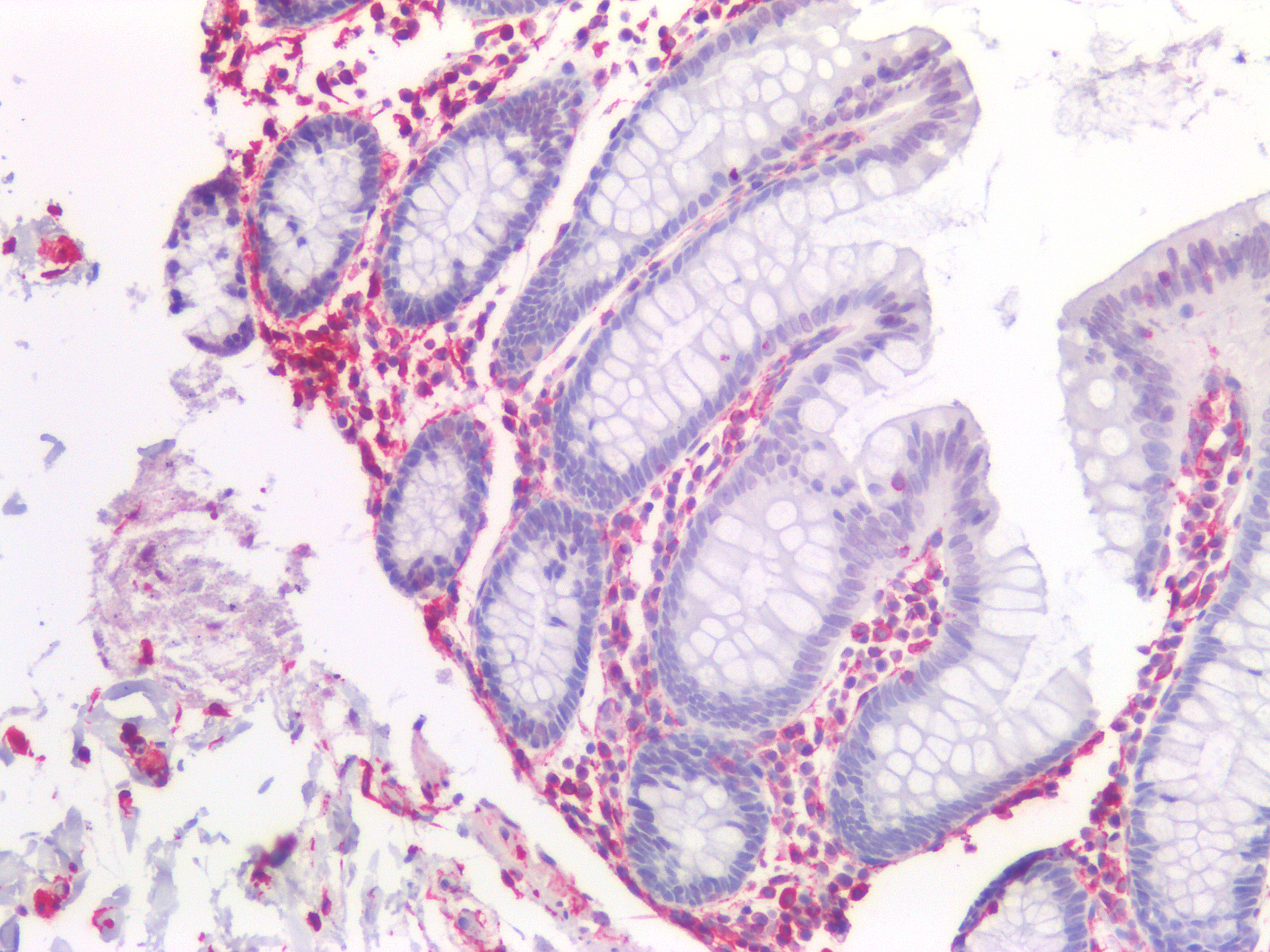
Figure 8. Immunostaining of human paraffin embedded tissue sections of human small intestine with MUB1903P (diluted 1:100), showing the specific pattern of vimentin in the mesenchymal cell types, such as fibroblasts in the connective tissue. As expected, no reactivity is seen in the epithelial cell compartment. |
|
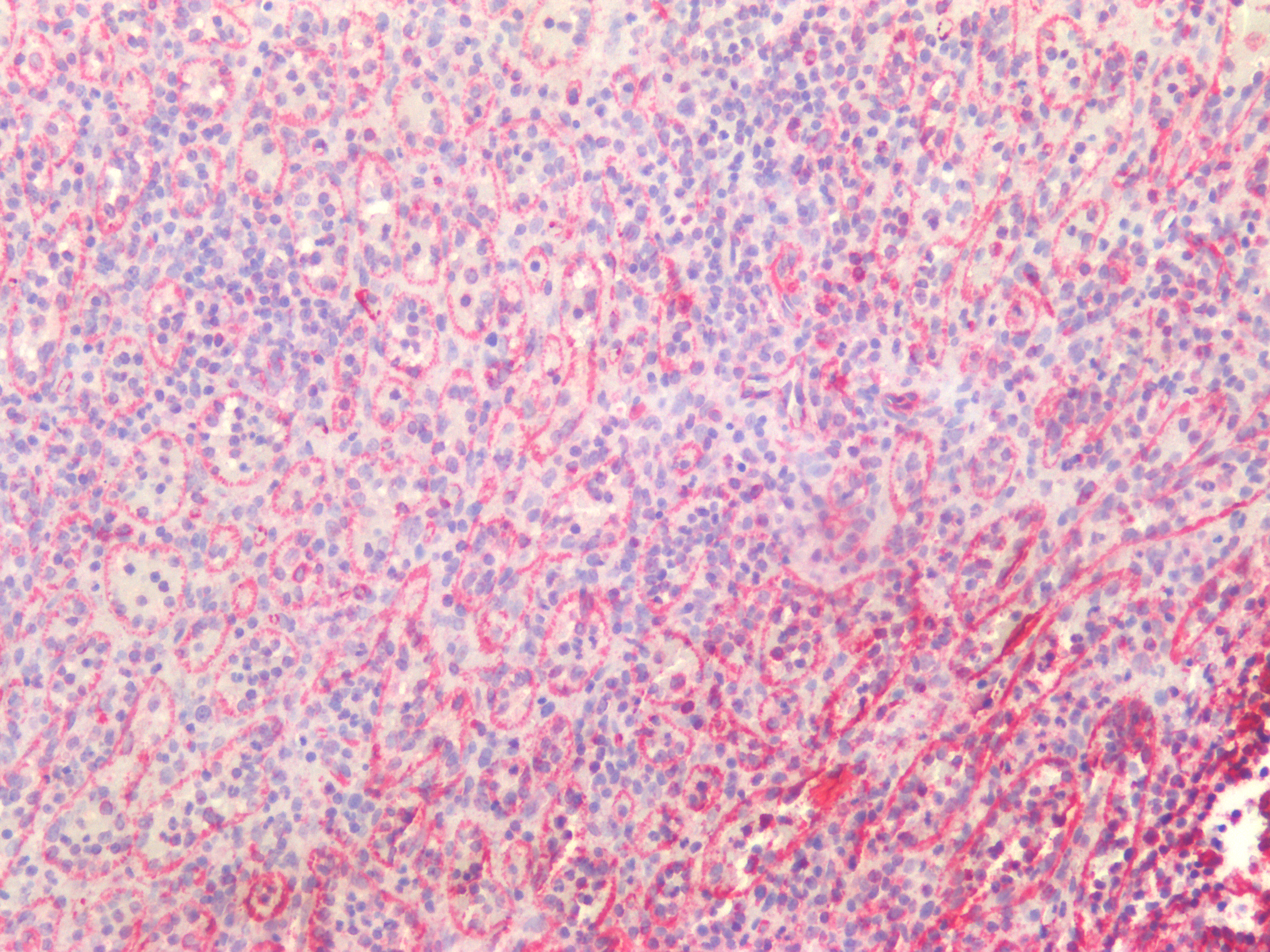
Figure 9. Immunostaining of human paraffin embedded tissue sections of human spleen with MUB1903P (diluted 1:100), showing the specific pattern of vimentin in the mesenchymal cell types. |
|
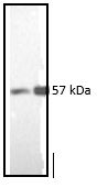
Figure 10. Western blotting result showing the specific reactivity of MUB1903P with the 57kDa protein band of vimentin in both the mouse (3T3 mouse fibroblasts; left lane) and human (normal human dermal fibroblasts; right lane) cell extracts. |
|
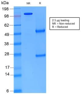
Figure 11. Non-reduced and reduced SDS-PAGE of MUB1903P showing its purity. |

Figure 1. Indirect immunofluorescence staining of normal human dermal fibroblasts in tissue culture with MUB1903P (diluted 1:250), showing the specific cytoskeletal pattern of vimentin intermediate filaments.

Figure 2. Indirect immunofluorescence staining of human kidney tissue section with MUB1903P (diluted 1:1000), showing the specific pattern of vimentin in the mesenchymal cell types, such as fibroblasts in the connective tissue, podocytes, and endothelial cells in blood vessels. As expected, no reactivity is seen in the epithelial cell compartment

Figure 3. Indirect immunofluorescence staining of human kidney tissue section with MUB1903P (diluted 1:1000), showing the specific pattern of vimentin in the mesenchymal cell types, such as fibroblasts in the connective tissue, podocytes, and endothelial cells in blood vessels. As expected, no reactivity is seen in the epithelial cell compartment

Figure 4. Immunostaining of human paraffin embedded tissue section of placenta with MUB1903P (diluted 1:100), showing the specific pattern of vimentin in the mesenchymal cell types, such as fibroblasts in the connective tissue, and endothelial cells in blood vessels. As expected, no reactivity is seen in the epithelial cell compartment

Figure 5. Immunostaining of human paraffin embedded tissue section of testis with MUB1903P (diluted 1:100), showing the specific pattern of vimentin in the mesenchymal cell types, such as fibroblasts in the connective tissue, endothelial cells in blood vessels and Sertoli cells.

Figure 6. Immunostaining of human paraffin embedded tissue sections of human colon with MUB1903P (diluted 1:100), showing the specific pattern of vimentin in the mesenchymal cell types, such as fibroblasts in the connective tissue, and endothelial cells in blood vessels. As expected, no reactivity is seen in the epithelial cell compartment.

Figure 7. Immunostaining of human paraffin embedded tissue sections of human kidney with MUB1903P (diluted 1:100), showing the specific pattern of vimentin in the mesenchymal cell types, such as fibroblasts in the connective tissue, and podocytes. As expected, no reactivity is seen in the epithelial cell compartment.

Figure 8. Immunostaining of human paraffin embedded tissue sections of human small intestine with MUB1903P (diluted 1:100), showing the specific pattern of vimentin in the mesenchymal cell types, such as fibroblasts in the connective tissue. As expected, no reactivity is seen in the epithelial cell compartment.

Figure 9. Immunostaining of human paraffin embedded tissue sections of human spleen with MUB1903P (diluted 1:100), showing the specific pattern of vimentin in the mesenchymal cell types.

Figure 10. Western blotting result showing the specific reactivity of MUB1903P with the 57kDa protein band of vimentin in both the mouse (3T3 mouse fibroblasts; left lane) and human (normal human dermal fibroblasts; right lane) cell extracts.

Figure 11. Non-reduced and reduced SDS-PAGE of MUB1903P showing its purity.