Catalogue

Mouse anti Neu-Oncogen (C-erb B2)
Catalog number: MUB1319P| Clone | 3B5 |
| Isotype | IgG1 |
| Product Type |
Primary Antibodies |
| Units | 0.1 mg |
| Host | Mouse |
| Species Reactivity |
Human Monkey Mouse Rat |
| Application |
Immunohistochemistry (frozen) Immunohistochemistry (paraffin) Immunoprecipitation Western Blotting |
Background
C-erbB-2 (erythroblastosis oncogene B), also known as HER2 (Human Epidermal Growth Factor Receptor 2), or Neu, CD340 and p185 is a protein that in Humans is encoded by the ERBB2 gene. AmplifiCation or overexpression of this gene has been shown to play an important role in the pathogenesis and progression of certain aggressive types of breast cancer, as well as many other epithelial malignancies and brain tumors. In recent years it has become an important biomarker and target of therapy for disease. ERBB2 is a known proto-oncogene loCated at the long arm of Human chromosome 17 (17q21-q22). The oncogene was found to code for EGFR. Gene cloning showed that HER2, Neu and ErbB-2 are all encoded by the same gene. The ErbB family is composed of plasma membrane-bound receptor tyrosine kinases, that contain an extracellular ligand binding domain, a transmembrane domain and an intracellular domain that can interact with a multitude of signaling molecules. HER2 can heterodimerise with any of the other three receptors and is considered to be the preferred dimerisation partner of the other ErbB receptors. Dimerisation results in the autophosphorylation of tyrosine residues within the cytoplasmic domain of the receptors and initiates a variety of signaling pathways.
Source
3B5 is a Mouse monoclonal IgG1 antibody derived by fusion of SP2/0 Mouse myeloma cells with spleen cells from a BALB/c Mouse immunized with a synthetic peptide TAENPEYLGLDVPV corresponding to amino acid residues 1242-1255 of the C-terminus of the Human c-erbB-2/HER-2/neu protein. This sequence is identical in Rat neu protein.
Product
Each vial contains 100 ul 1 mg/ml purified monoclonal antibody in PBS containing 0.09% sodium azide.
Formulation: Each vial contains 100 ul 1 mg/ml purified monoclonal antibody in PBS containing 0.09% sodium azide.
Specificity
3B5 reacts equally well with the wild type as well as the mutant (oncogenic) form of the c-erbB-2/HER-2/neu protein, but preferentially recognizes the unphosphorylated form of this protein.
Applications
3B5 is useful for immunohistochemistry on frozen and paraffin-embedded tissues, immunocytochemistry, flow cytometry, immunoprecipitation and immunoblotting. Optimal antibody dilution should be determined by titration. Recommended range is 1:100 – 1:500 for immunohistochemistry and 1:250 – 1:1000 for immunoblotting applications. For appliCation on paraffin embedded tissue antigen retrieval by boiling in 10mM citRate buffer pH 6.0 is recommended.
Storage
The antibody is shipped at ambient temperature and may be stored at +4°C. For prolonged storage prepare appropriate aliquots and store at or below -20°C. Prior to use, an aliquot is thawed slowly in the dark at ambient temperature, spun down again and used to prepare working dilutions by adding sterile phosphate buffered saline (PBS, pH 7.2). Repeated thawing and freezing should be avoided. Working dilutions should be stored at +4°C, not refrozen, and preferably used the same day. If a slight precipitation occurs upon storage, this should be removed by centrifugation. It will not affect the performance or the concentration of the product.
Caution
This product is intended FOR RESEARCH USE ONLY, and FOR TESTS IN VITRO, not for use in diagnostic or therapeutic procedures involving humans or animals. It may contain hazardous ingredients. Please refer to the Safety Data Sheets (SDS) for additional information and proper handling procedures. Dispose product remainders according to local regulations.This datasheet is as accurate as reasonably achievable, but Nordic-MUbio accepts no liability for any inaccuracies or omissions in this information.
References
1. Van de Vijver, M.J., Peterse, J.L., Mooi, W.J., Wisman, P., Lomans, J., Dalesio, O. and Nusse, R. (1988). Neu-protein overexpression in breast cancer. Association with comedo-type ductal carcinoma in situ and limited prognostic value in stage II breast cancer.. N Engl J Med. 319, 1239-45.
2. De Potter, C.R., Van Daele, S., Van de Vijver, M.J., Pauwels, C., Maertens, G., De Boever, J., Vandekerckhove, D. and Roels, H. (1989). The expression of the neu oncogene product in breast lesions and in normal fetal and adult Human tissues. Histopathology 15, 351-62.
3. De Potter, C.R., Quatacker, J., Maertens, G., Van Daele, S., Pauwels, C., Verhofstede, C., Eechaute, W. and Roels, H. (1989). The subcellular localization of the neu protein in Human normal and neoplastic cells. Int J Cancer 44, 969-74.
4. Van Leeuwen, F., Van de Vijver, M.J., Lomans, J., Van Deemter, L., Jenster, G., Akiyama, T., Yamamoto, T. and Nusse, R. (1990). Mutation of the Human neu protein facilitates down-modulation by monoclonal antibodies. Oncogene. 5, 497-503.
6. Singleton, T.P., Niehans, G.A., Gu, F., Litz, C.E., Hagen, K., Qiu, Q., Kiang, D.T. and Strickler, J.G. (1992). Detection of c-erbB-2 activation in paraffin-embedded tissue by immunohistochemistry. Hum Pathol. 23, 1141-50.
7. Schwechheimer, K., Läufle, R.M., Schmahl, W., Knödlseder, M., Fischer, H. and Höfler, H. (1994). Expression of neu/c-erbB-2 in Human brain tumors. Hum Pathol. 25, 772-80.
Safety Datasheet(s) for this product:
| NM_Sodium Azide |

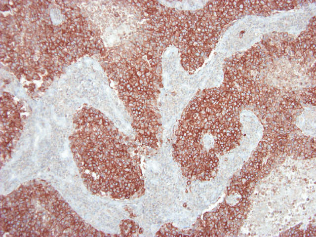
Figure 1. Immunohistochemistry of MUB1319P on formalin fixed, paraffin embedded tissue section of human breast carcinoma. Dilution 1:100.

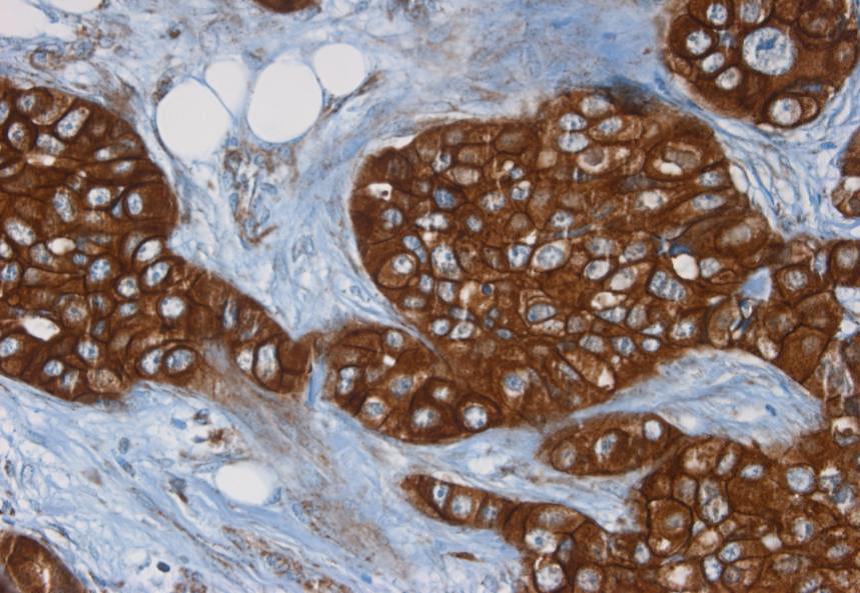
Figure 2. Immunohistochemistry of MUB1319P on formalin fixed, paraffin embedded tissue section of human breast carcinoma. Dilution 1:100.

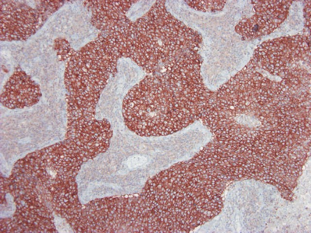
Figure 3. Immunohistochemistry of MUB1319P on formalin fixed, paraffin embedded tissue section of human breast carcinoma. Dilution 1:1000.


