Catalogue

Mouse anti NCAM / CD56
Catalog number: MUB1301P| Clone | 123C3 |
| Isotype | IgG1 |
| Product Type |
Primary Antibodies |
| Units | 0.1 mg |
| Host | Mouse |
| Species Reactivity |
Human Zebrafish |
| Application |
Immunocytochemistry Immunohistochemistry (frozen) Immunohistochemistry (paraffin) Western Blotting |
Background
NCAM, as a member of the immunoglobulin superfamily of adhesion molecules is characterized by several immunoglobulin (Ig)-like domains. The extracellular part of NCAM consists of five of these Ig domains and two fibronectin type III homology regions. NCAM is encoded by a single copy gene composed of 26 exons. However, at least 20-30 distinct isoforms can be geneRated by alternative splicing and by posttranslational modifiCations, such as sialylation. During sialylation, polysialic acid (PSA) carbohydRates are attached to the extracellular part of NCAM. Through its extracellular region, NCAM mediates homophilic interactions. In addition, NCAM can also undergo heterophilic interactions by binding extracellular matrix components, such as laminin, or other cell adhesion molecules, such as integrins.
Source
123C3 is a Mouse monoclonal IgG1 antibody derived by fusion of Mouse myeloma cells with spleen cells from a Mouse immunized with the a membrane preparation of a small cell lung carcinoma specimen.
Product
Each vial contains 100 ul 1 mg/ml purified monoclonal antibody in PBS containing 0.09% sodium azide.
Formulation: Each vial contains 100 ul 1 mg/ml purified monoclonal antibody in PBS containing 0.09% sodium azide.
Specificity
123C3 was defined as a cluster I antibody during the First International Workshop on Small Cell Lung Cancer (SCLC) Antibodies. 123C3 recognizes an epitope in the NCAM exons 11-13 which is dependent on an intact conformation of the first fibronectin type-III homologous domain encoded by these exons. 123C3 stains NCAM which is present in small cell lung cancer and lung carcinoids. It also reacts with a number of non-small cell lung carcinomas and neuroendocrine and neuronal derived tissues. In addition, 123C3 is internalized after binding to its antigen on SCLC cell lines, making it an excellent reagent for tumor imaging in xenograft models.
Applications
123C3 is suitable for immunoblotting, immunocytochemistry and immunohistochemistry on frozen and paraffin-embedded tissues. Optimal antibody dilution should be determined by titration; recommended range is 1:100 – 1:200 for immunohistochemistry with avidin-biotinylated Horseradish peroxidase complex (ABC) as detection reagent, and 1:100 – 1:1000 for immunoblotting applications.
Storage
The antibody is shipped at ambient temperature and may be stored at +4°C. For prolonged storage prepare appropriate aliquots and store at or below -20°C. Prior to use, an aliquot is thawed slowly in the dark at ambient temperature, spun down again and used to prepare working dilutions by adding sterile phosphate buffered saline (PBS, pH 7.2). Repeated thawing and freezing should be avoided. Working dilutions should be stored at +4°C, not refrozen, and preferably used the same day. If a slight precipitation occurs upon storage, this should be removed by centrifugation. It will not affect the performance or the concentration of the product.
Caution
This product is intended FOR RESEARCH USE ONLY, and FOR TESTS IN VITRO, not for use in diagnostic or therapeutic procedures involving humans or animals. It may contain hazardous ingredients. Please refer to the Safety Data Sheets (SDS) for additional information and proper handling procedures. Dispose product remainders according to local regulations.This datasheet is as accurate as reasonably achievable, but Nordic-MUbio accepts no liability for any inaccuracies or omissions in this information.
References
1. Schol, D. J., Mooi, W. J., van der Gugten, A. A., Wagenaar, S. S., and Hilgers, J. (1988). Monoclonal antibody 123C3, identifying small cell carcinoma phenotype in lung tumours, recognizes mainly, but not exclusively, endocrine and neuron-supporting normal tissues, Int J Cancer Suppl 2, 34-40.
2. Mooi, W. J., Wagenaar, S. S., Schol, D., and Hilgers, J. (1988). Monoclonal antibody 123C3 in lung tumour classifiCation. Immunohistology of 358 resected lung tumours, Mol Cell Probes 2, 31-7.
3. Moolenaar, C. E., Muller, E. J., Schol, D. J., Figdor, C. G., Bock, E., Bitter-Suermann, D., and Michalides, R. J. (1990). Expression of neural cell adhesion molecule-related sialoglycoprotein in small cell lung cancer and neuroblastoma cell lines H69 and CHP-212, Cancer Res 50, 1102-6.
4. Kibbelaar, R. E., Moolenaar, K. E., Michalides, R. J., Van Bodegom, P. C., Vanderschueren, R. G., Wagenaar, S. S., Dingemans, K. P., Bitter-Suermann, D., Dalesio, O., Van Zandwijk, N., and et al. (1991). Neural cell adhesion molecule expression, neuroendocrine differentiation and prognosis in lung carcinoma, Eur J Cancer 27, 431-5.
5. Gerardy-Schahn, R., and Eckhardt, M. (1994). Hot spots of antigenicity in the neural cell adhesion molecule NCAM, Int J Cancer Suppl 8, 38-42.
6. Kwa, H. B., Verhoeven, A. H., Storm, J., van Zandwijk, N., Mooi, W. J., and Hilkens, J. (1995). Radioimmunotherapy of small-cell lung cancer xenografts using 131I- labelled anti-NCAM monoclonal antibody 123C3, Cancer Immunol Immunother 41, 169-74.
7. Kwa, H. B., Verheijen, M. G., Litvinov, S. V., Dijkman, J. H., Mooi, W. J., and Van Krieken, J. H. (1996). Prognostic factors in resected non-small cell lung cancer: an immunohistochemical study of 39 cases, Lung Cancer 16, 35-45.
Safety Datasheet(s) for this product:
| NM_Sodium Azide |

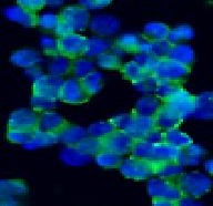
Figure 1. Immunostaining of the neural cell adhesion molecule NCAM/CD56 in the small cell lung cancer cell line NCI-H82 using MUB1301P.

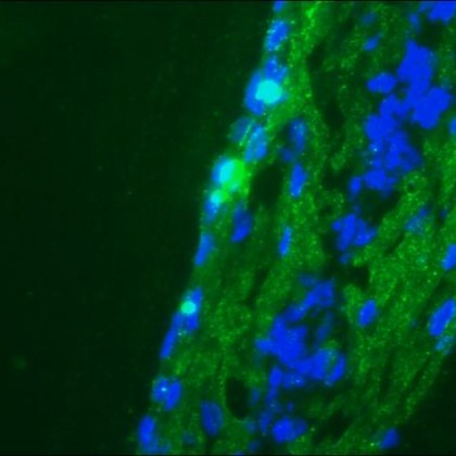
Figure 2. Immunofluorescence staining of a 9-days-old zebrafish-embryo using MUB1301P.


