Catalogue

Mouse anti actin alpha-muscle
Catalog number: MUB0107P| Clone | HHF35 |
| Isotype | IgG1 |
| Product Type |
Primary Antibodies |
| Units | 0.1 mg |
| Host | Mouse |
| Species Reactivity |
Chicken Human Monkey Rabbit Rat Swine Zebrafish |
| Application |
Electron microscopy ELISA Immunocytochemistry Immunohistochemistry (frozen) Immunohistochemistry (paraffin) Western Blotting |
Background
Among the six actin isoforms described in mammals, two are found in virtually all cells (β- and γ-cytoplasmic), two are detected in smooth muscle cells (α- and γ-smooth muscle) and two are present in striated muscles, one predominantly in skeletal (α-skeletal) and one in cardiac (α-cardiac) muscle cells. These actin isoforms differ slightly in their N-terminus, but the sequence of each of these actins is highly conserved in higher vertebRates. Alpha- muscle actin is present in striated as well as smooth muscle cells, and in pathological tissues derived therefrom. It has for example been detected in several types of muscle derived tumors, and also been shown to appear in stress fibers of fibroblastic cells involved in contractile phenomena such as wound healing and fibrocontractive diseases.
Source
HHF35 is a Mouse monoclonal IgG1 antibody derived by fusion of NS-1 Mouse myeloma cells with spleen cells from a BALB/c Mouse immunized with an SDS-extracted protein fraction from Human myocardium.
Product
Each vial contains 50 ul 1 mg/ml purified monoclonal antibody in PBS containing 0.09% sodium azide.
Formulation: Each vial contains 50 ul 1 mg/ml purified monoclonal antibody in PBS containing 0.09% sodium azide.
Specificity
HHF35 reacts with both α-muscle and γ-smooth muscle actin, and therefore reacts with skeletal muscle, cardiac muscle, vascular and visceral smooth muscle cells, pericytes and myoepithelial cells. It is also reactive in myofibroblasts. It does not react with epithelial, endothelial, neural or normal connective tissue cells when applied under the proper conditions to these tissue sections.
Species Reactivity: The epitope recognized by α-SM1 is highly conserved. The antibody therefore cross-reacts with many species including protochordates, lower craniates and mammals.
Applications
HHF35 is useful for immunohistochemistry on frozen and paraffin-embedded tissues preserved in several types of fixatives (see reference 1), immunoblotting, immuno-electron microscopy and ELISA. For immunohistochemical applications to paraffin embedded tissues it is recommended to dilute the antibody in PBS containing 50 mM EDTA. Optimal antibody dilution should be determined by titration; recommended range is 1:200 – 1:1000 for immunohistochemistry with avidin-biotinylated Horseradish peroxidase complex (ABC) as detection reagent, and 1:1000 – 1:5000 for immunoblotting applications.
Storage
The antibody is shipped at ambient temperature and may be stored at +4°C. For prolonged storage prepare appropriate aliquots and store at or below -20°C. Prior to use, an aliquot is thawed slowly in the dark at ambient temperature, spun down again and used to prepare working dilutions by adding sterile phosphate buffered saline (PBS, pH 7.2). Repeated thawing and freezing should be avoided. Working dilutions should be stored at +4°C, not refrozen, and preferably used the same day. If a slight precipitation occurs upon storage, this should be removed by centrifugation. It will not affect the performance or the concentration of the product.
Caution
This product is intended FOR RESEARCH USE ONLY, and FOR TESTS IN VITRO, not for use in diagnostic or therapeutic procedures involving humans or animals. It may contain hazardous ingredients. Please refer to the Safety Data Sheets (SDS) for additional information and proper handling procedures. Dispose product remainders according to local regulations.This datasheet is as accurate as reasonably achievable, but Nordic-MUbio accepts no liability for any inaccuracies or omissions in this information.
References
1. Tsukada, T., Tippens, D., Gordon, D., Ross, R. and Gown, A.M. (1987). HHF35, a muscle-actin-specific monoclonal antibody. I. Immunocytochemical and biochemical characterization. American Journal of Pathology 126, 51-60.
2. Tsukada, T., McNutt, M.A., Ross, R. and Gown, A.M. (1987). HHF35, a muscle-actin-specific monoclonal antibody. II. Reactivity in normal, reactive, and neoplastic Human tissues. American Journal of Pathology 127, 389-402.
3. Schmidt, R.A., Cone, R., Haas, J.E. and Gown, A.M. (1988). Diagnosis of rhabdomyosarcomas with HHF35, a monoclonal antibody directed anti muscle actins. American Journal of Pathology 131, 19-28.
4. Babaev, V.R., Bobryshev, Y.V., Stenina, O.V., Tararak, E.M. and Gabani, G. (1990). Heterogeneity of smooth muscle in atheromatous plaque of Human aorta. American Journal of Pathlogy 136, 1031-42.
5. Nascimento, C., Caroli-Bottino, A., Paschoal, J. and Pannain, V.L. (2009). Vascular immunohistochemical markers: contributions to hepatocellular nodule diagnosis in explanted livers. Transplantation Proceedings 41, 4211-13.
Protein Reference(s)
Database Name: UniProt
Accession Number: P62736 & P68032
Safety Datasheet(s) for this product:
| NM_Sodium Azide |

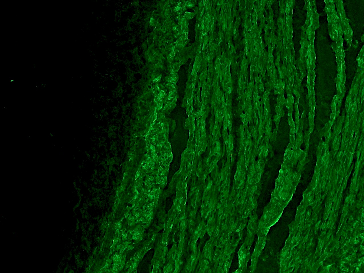
Figure 1. Indirect immunofluorescence staining of frozen section of chicken gizzard with MUB0107P (HHF35) showing specific positive staining of smooth muscle cells.

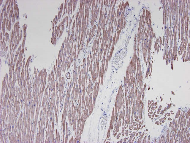
Figure 2. Formalin fixed, paraffin embedded human heart tissue immunostained for actin with MUB0107P (clone HHF35) at a 1:250 dilution.

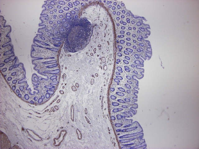
Figure 3. Formalin fixed, paraffin embedded human small intestine, immunostained for actin using MUB0107P (clone HHF35) at a 1:100 dilution. Note staining of smooth muscle cells and no reactivity on the epithelium and connective tissue.

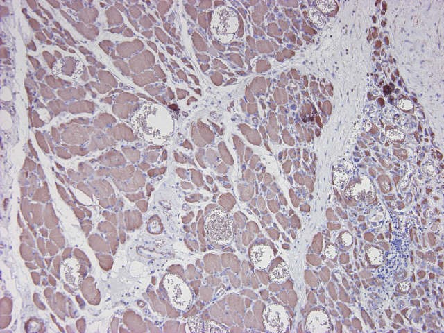
Figure 4. Formalin fixed, paraffin embedded human tongue tissue, immunostained for actin using MUB0107P (clone HHF35) at a 1:250 dilution. Note staining of striated muscle cells and no reactivity on the epithelium and connective tissue.


