Catalogue

BrdU Immunohistochemistry Kit
Catalog number: X1545K.1| Product Type |
Kits |
| Units | 50 Slides |
| Application |
Immunocytochemistry Immunohistochemistry (frozen & paraffin) |
Background
The BrdU Immunohistochemistry Kit is a non-isotopic enzyme immunoassay for the detection and quantification of DNA synthesis and cell proliferation. Evaluation of cell cycle progression is essential for investigations in many scientific fields. Measurement of [3H] thymidine incorporation as cells enter S-phase has long been the traditional method for the detection of cell proliferation. Subsequent quantification of [3H] thymidine is performed by scintillation counting or autoradiography. This technology is slow, labor intensive and has several limitations including the handling and disposal of radioisotopes and the necessity of expensive equipment. A well-established alternative to [3H] thymidine uptake has been demonstrated by numerous investigators. In these methods bromodeoxyuridine (BrdU), a thymidine analog, replaces [3H] thymidine. BrdU is incorporated into newly synthesized DNA strands of actively proliferating cells. Following partial denaturation of double stranded DNA, BrdU is detected immunochemically allowing the assessment of the population of cells which are actively synthesizing DNA. Exalpha Biologicals BrdU Immunohistochemistry Kit involves incorporation of BrdU Reagent into cells, either in tissues after injection of BrdU in experimental animals or in cells cultured in microtiter plates or on glass slides. The resultant assay is, rapid, easy to perform and applicable to high sample throughput. In addition to evaluation of cell proliferation, information such as cell number, morphology and analysis of cellular antigens can be obtained from a single culture or tissue section.
Product
The BrdU Cell Immunohistochemistry Kit contains all ingredients (antibodies, buffers and detection reagents) needed for the immunohistochemical detection and quantification of the proliferative activity of cells and tissues. The kit contains 5 control slides with BrdU prelabeled MCF-7 cells (one of which is prestained) as well as an instruction manual describing the relevant protocols for BrdU immunostaining.
Applications
Immunohistochemistry on formalin fixed, paraffin embedded, as well as frozen tissue sections. Furthermore this kit can be used for the immunocytochemical staining of cultured cells on glass slides or cell culture plates. Optimal concentration of the primary antibody should be evaluated by serial dilutions.
Storage
Upon receipt, store entire kit at -20°C. Once the kit is thawed, you may keep it at 4°C for 5 days. For long-term storage, it is recommended you aliquot and freeze the components at –20°C, particularly the Streptavidin-HRP Conjugate, the Prediluted Biotinylated Sheep anti-BrdU Detector Antibody and the 4x Trypsin Concentrate.
Shipping Conditions: The Exalpha Biologicals BrdU Immunohistochemistry Kit components are shipped on blue ice.
Caution
This product is intended FOR RESEARCH USE ONLY, and FOR TESTS IN VITRO, not for use in diagnostic or therapeutic procedures involving humans or animals. It may contain hazardous ingredients. Please refer to the Safety Data Sheets (SDS) for additional information and proper handling procedures. Dispose product remainders according to local regulations.This datasheet is as accurate as reasonably achievable, but Nordic-MUbio accepts no liability for any inaccuracies or omissions in this information.
Safety Datasheet(s) for this product:
| EA_X1545K.1-SDS_V1 |
Kit Manual
Click here to download the Kit Manual

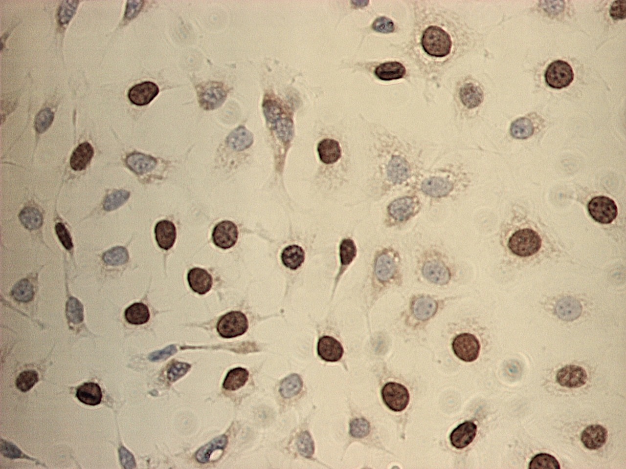
Figure 1: BrdU labeled MCF-7 cells immunostained using the X1545K.1 kit. Note the specific nuclear staining of proliferating S-phase cells.

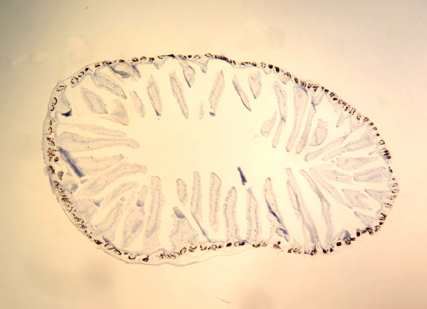
Figure 2: Immunolabeling of mouse small intestine with the X1545K.1 kit showing BrdU positive cells in the proliferating compartment of the crypts (low magnification).

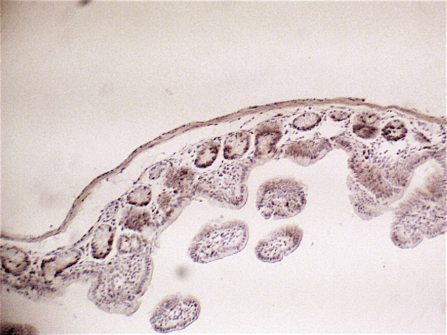
Figure 3: Immunolabeling of mouse small intestine with the X1545K.1 kit showing BrdU positive cells in the proliferating compartment of the crypts (high magnification)

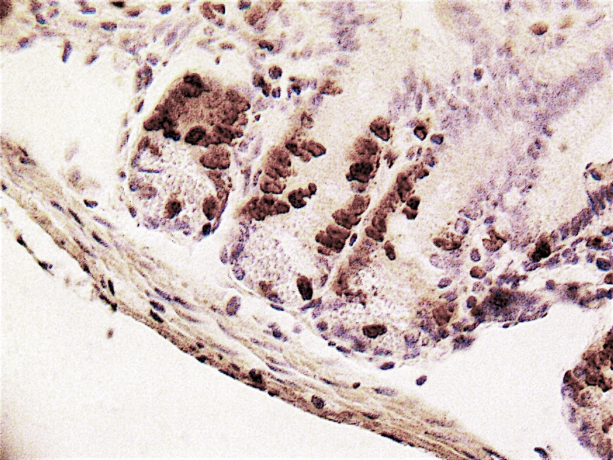
Figure 4: Immunolabeling of mouse small intestine with the X1545K.1 kit showing BrdU positive cells in the proliferating compartment of the crypts (high magnification

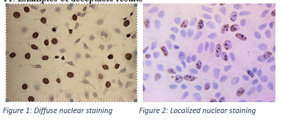
Figure 5. MCF-7 cells stained for BrdU incorparation using the X1545K.1 kit. Note the exclusive nuclear immunostaining with a diffuse staining pattern (Fig. 1) and a more localized pattern (Fig. 2).

